When your blot looks noisy or your fluorescent image fades, don’t blame the primary antibody. The real culprit is often the secondary. ICL shows you how to choose wisely and watch faint bands pop clear.
When a blot looks noisy, a fluorescent image fades, or an ELISA misses the low end of the curve, we usually blame the primary antibody. Yet in most troubleshooting sessions the real issue is the secondary. Pick the wrong one and you amplify every bit of background. When chosen correctly faint band pops into crystal-clear view. Below is a friendly guide to why secondaries matter and how to make them work for you.
1. What a Secondary Antibody Actually Does
Boosts signal. One primary antibody binds a single epitope. Several secondaries can bind that primary, stacking enzymes or fluorophores for a much stronger read-out.
Adds flexibility. Change the label on your secondary and you can move the same primary from a blot to an ELISA or a rapid flow assay without buying new primaries.
Saves money. Primaries are pricey. A well-chosen secondary lets one primary cover many applications.
2. Three quality checks that matter
Quality check
Why it counts
Quick way to confirm
Specificity
Less background, fewer mystery bands
Look for cross-adsorbed or highly adsorbed IgG.
Affinity or titer
Stronger signal at higher dilution
Review the supplier’s recommended range and QC data.
Label integrity
Consistent brightness or enzyme activity
Ask for fluorophore-to-protein ratio or HRP activity on the COA.
3. Matching the secondary to the job
Western blot
Goat anti-mouse or anti-rabbit IgG tagged with HRP is the workhorse. For very low abundance targets, an HRP polymer secondary often brings a solid order-of-magnitude boost.
ELISA and Lateral Flow
Background can occur when using serum and/or milk for diluents or blocking buffers. Cross-adsorbed HRP secondaries keep it in check. If you prefer an avidin-biotin system, switch to streptavidin-HRP plus a biotinylated secondary to avoid endogenous biotin issues.
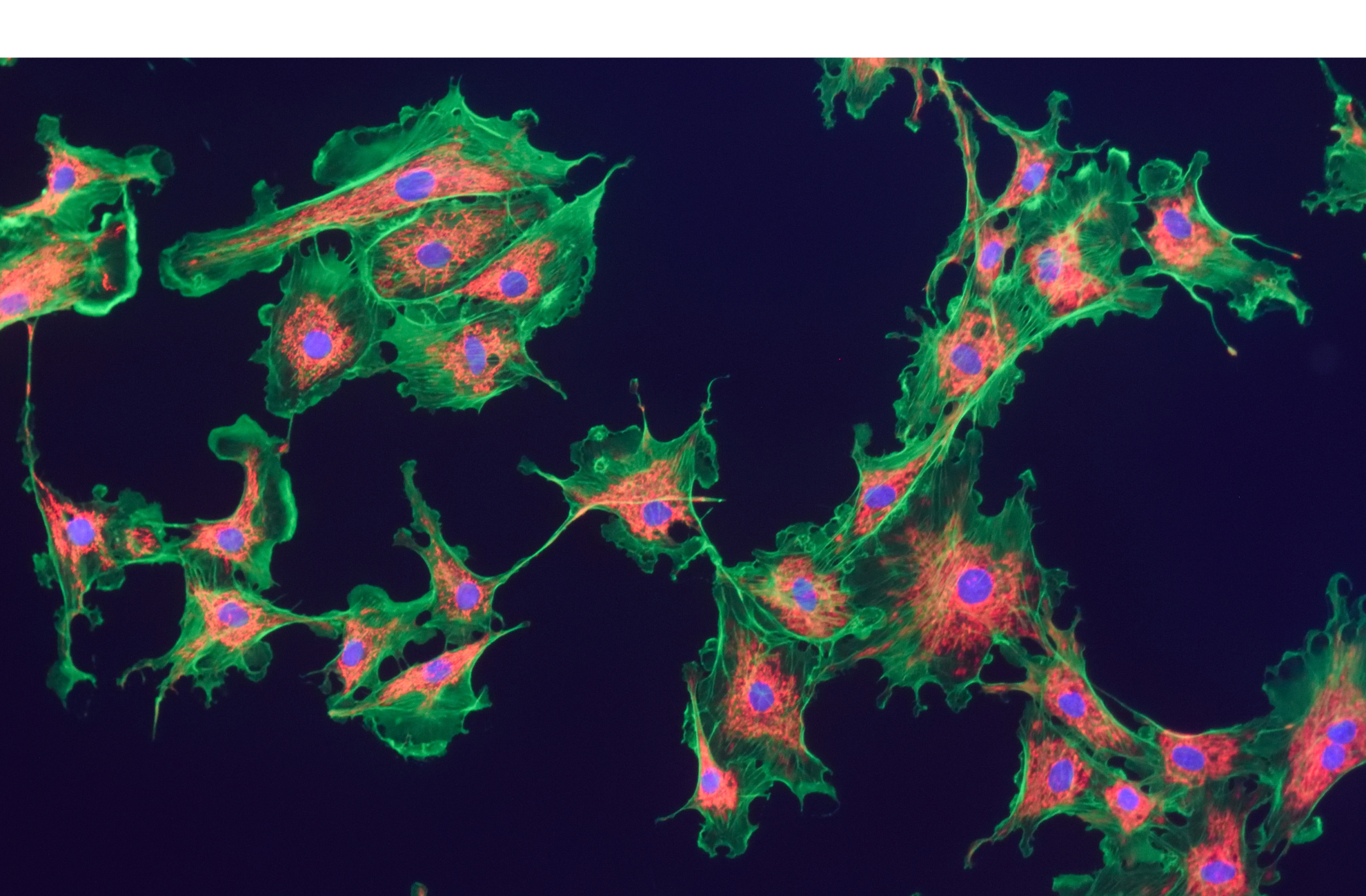
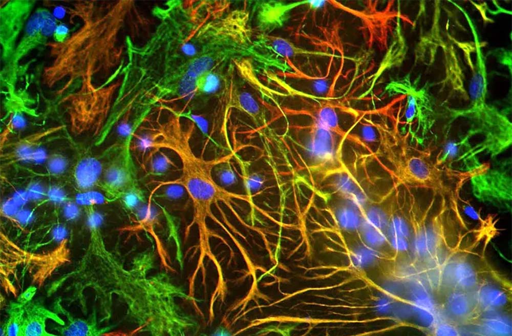
Immunofluorescence and flow cytometry
Brightness and photostability rule. Far-red dyes like Alexa Fluor 647 or CF 647 cut tissue autofluorescence and stay bright longer than classic FITC.
4. Common Pitfalls and How to Dodge Them
Using stock concentration. Start with a checkerboard titration. Often a 1 in 5,000 dilution gives cleaner data than 1 in 1,000 and saves reagent.
Host mismatch. If your primary is mouse and your tissue is also mouse, consider a Fab fragment or subclass-specific secondary to avoid endogenous IgG.
Light damage. For IF, protect slides from light and choose dyes with high photostability.
Lot drift. Secure a single lot for long studies or regulated work to keep your signal consistent.
5. Looking Ahead
Recombinant secondaries remove batch variability.
Nanobody-based secondaries shrink steric hindrance, ideal for super-resolution imaging.
DNA barcoded secondaries allow hybridization chain reactions for huge signal gains.
6. Quick Checklist Before you Hit “Order”
Match host species- Secondary antibodies are produced in several host species, including goat, rabbit, donkey, and chicken. Always pick a secondary raised in a species different from your primary antibody host. For example, if the primary antibody is rabbit, select an anti-rabbit secondary made in goat, donkey, or chicken instead of rabbit.
Confirm cross-adsorption- Pre-absorbed secondary antibodies have been screened to remove any unwanted cross-reactivity with specific animal species. Using a secondary that is pre-adsorbed against the species in your sample keeps it from binding the wrong proteins. For instance, when working with human tissue you may want a secondary that does not react with human proteins, otherwise you risk false positives or a noisy background.
Pick the right conjugate - Match the conjugate to the assay. In Western blots and ELISAs, a secondary antibody tagged with horseradish peroxidase, alkaline phosphatase, or biotin works best. A biotinylated secondary used together with a labeled streptavidin, such as streptavidin alkaline phosphatase or streptavidin HRP, can increase the signal by about four times.
Run a dilution series to nail the best signal-to-noise.
Reserve enough of one lot for the life of the project.
Ready to upgrade?
Immunology Consultants Laboratory (ICL) offers classic HRP / Biotin / FITC conjugated secondaries along with unconjugated antibodies. All come with QC certified and technical support that answers within a day.
Antibodies | ELISA | Proteins | Veterinary and Human ELISA Kits | Host Cell Protein (HCP) ELISA Kits | WB | Lateral Flow Immunoassay Reagents | R&D to IVD